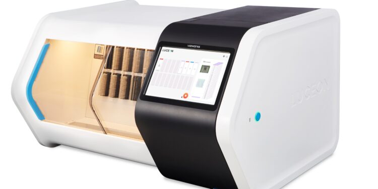
Vieworks, a leading name in bioimaging solutions, is to set a new standard in the digital pathology field with the launch of its high-speed slide scanner LUCEON 510. Vieworks will be making its debut as a digital pathology solution provider at the upcoming European Congress of Pathology (ECP) 2024, one of the most prestigious events in the digital pathology community, in Florence, Italy (Booth 39, September 7th – 10th).
Employing the intricately designed 3-camera structure, LUCEON delivers four different imaging modes each optimized for varying applications, including histopathology slides, liquid-based cytology slides, and cytology slides. Three Vieworks cameras—developed and manufactured entirely by Vieworks—obtain three differently focused images at once with scan time and data size comparable to that of a single camera.
Among the four imaging modes, the tri-focal layer merge mode captures and fuses three individual images real-time, resulting in a single fusion image containing the best focused pixels from each image. A variation of the conventional Z-stack, the tri-focal layer merge Z-stack mode utilizes LUCEON’s 3-camera structure to acquire images with a scan time 3 times faster and image size one-third of Z-stack.
LUCEON 510 loads up to 510 slides when fully loaded with 17 racks, 30 slides per rack. Vieworks’ patent pending compatibility adapter allows the use of both 30 slide racks and 20 slide racks. With a scan speed of 30 seconds per slide, LUCEON scans up to 83 slides per hour (15 mm x 15 mm, 40x).
Four Imaging Modes
- Best focused layer mode
– Automatically selects image with best image quality
– Histopathology slides - Tri-focal layer merge mode
– Three individual images are captured and fused real-time
– Resulting fusion image contains best focused pixels from each individual image
– Liquid-based cytology slides - Z-stack mode
– High speed Z-stack
– Adjusts layer spacing in increments of 0.1 ㎛
– Cytology slides - Tri-focal layer merge Z-stack
– High speed tri-focal layer merge Z-stack
– Adjusts layer spacing in increments of 0.1 ㎛ (recommended spacing: 1 ㎛)
– Cytology slides


